How the receptor is built
Immune cells do not drift toward a problem. They cut their way there, dissolving a path through the mesh of protein that holds tissue together. uPAR is the tool that lets them aim the cutting.
The urokinase plasminogen activator receptor sits on the outside of the cell membrane, held there by a lipid tail rather than passing through the membrane as most receptors do. Its three domains, D1, D2 and D3, form a concave shape with a cone-shaped cavity at the center, into which the business end of urokinase inserts (Huai, Science 2006). Its gene, PLAUR (Casey, Blood 1994), reappears later: a common variant in it sets how much of the soluble form a person carries.
What the receptor does is localize. Urokinase converts plasminogen into plasmin, an enzyme that cuts through the scaffolding between cells. Free in solution that would be indiscriminate. Docked to uPAR, it is concentrated at the point where the cell meets the scaffolding, so the cutting happens where the cell is actually trying to go (Appella, J Biol Chem 1987).
It is not only a dock for an enzyme. uPAR binds vitronectin, one of the proteins of that scaffolding, with high affinity and independently of urokinase, which makes it an adhesion receptor in its own right (Wei, J Biol Chem 1994). And because no part of it crosses into the cell, it cannot signal alone. It works by partnering, most consequentially with the integrin Mac-1 on monocytes, whose adhesive behavior it regulates (Simon, Blood 1996).
It appears when cells are called
Most receptors are furniture. This one is equipment: kept in store, brought out when it is needed, and put out in different amounts depending on what the cell is being told.
uPAR was first identified on monocytes as Mo3, an antigen that shows up when the cell is activated (Min, J Immunol 1992). Neutrophils keep it in two separate compartments inside the cell and move it to the surface on stimulation (Plesner, Blood 1994), so a cell can raise its own surface density within minutes without making any new protein. Over longer periods the gene itself is turned up, by interferon-γ, by tumor necrosis factor-α (Kirchheimer, J Immunol 1988) and by low oxygen acting through a separate pathway (Graham, Blood 1998).
The detail that matters most for anything measured in blood is that the two forms are not regulated together. In mononuclear phagocytes, interferon-γ raises surface uPAR two- to three-fold; tumor necrosis factor-α leaves the surface unchanged while raising the messenger RNA, the internal pool and the amount of receptor shed (Sitrin, Blood 1994). One signal builds the tool. Another releases the fragment. A rising blood level is therefore not a simple readout of how much receptor is on cells.
Where the circulating protein comes from is still being worked out, and the answer is not only mature blood cells: in chronic kidney disease, bone marrow shows proinflammatory reprogramming of monocyte precursors, and the myeloid cells this produces secrete suPAR (Spear, J Am Soc Nephrol 2026).
This is what makes the receptor worth measuring at all. A housekeeping protein reports on the machinery of ordinary life, and its level says little about a person. A protein a cell puts out only when it is called reports something else: how often, and how hard, that call has come. What accumulates in the blood is a record of that demand.
What it is for
Take the receptor away and immune cells are still made, but they arrive late, or not at all.
In human cells, blocking uPAR with an antibody cuts monocyte movement toward a chemical signal to 14.2% of untreated cells, a level the authors found indistinguishable from random drift. Blocking urokinase itself does almost nothing, leaving 88.2% of normal movement intact (Gyetko, J Clin Invest 1994). What the cell needs is the receptor, not the enzyme it carries.
The same holds in a living animal. Mice without uPAR are healthy and fertile at rest but bring roughly half as many white cells into an inflamed abdomen, with granulocyte recruitment nearly abolished (Bugge, J Biol Chem 1995; May, J Exp Med 1998), and against Pseudomonas aeruginosa pneumonia they fail to bring neutrophils into the lung and clear the infection poorly, while mice lacking urokinase recruit indistinguishably from normal (Gyetko, J Immunol 2000). No inherited uPAR deficiency has ever been described in a person, so how far this carries to humans is genuinely unknown.
How the soluble form is made
suPAR is the receptor without its anchor. There are two ways it gets that way, and they do not produce the same molecule.
The lipid anchor can be cut, releasing the whole three-domain receptor into the circulation. Or the receptor can be cleaved between D1 and D2, by urokinase itself or by plasmin, releasing a two-domain fragment and a free D1 (Høyer-Hansen, J Biol Chem 1992; Sidenius, FEBS Lett 2000).
The second route is not disposal. It is a change of job. Removing D1 abolishes the receptor's association with integrins and its ability to regulate adhesion (Montuori, J Biol Chem 2002), and it exposes a short sequence that acts on FPRL1, a receptor cells use to follow chemical gradients, so the fragment becomes a signal that draws other immune cells in (Resnati, Proc Natl Acad Sci U S A 2002). Activated neutrophils release it rapidly (Pliyev, Mol Cell Biochem 2009).
So what a laboratory reports as suPAR is a mixture of forms with different biology, and assays that recognize different parts of the molecule do not measure quite the same thing.
What the soluble form does that the receptor cannot
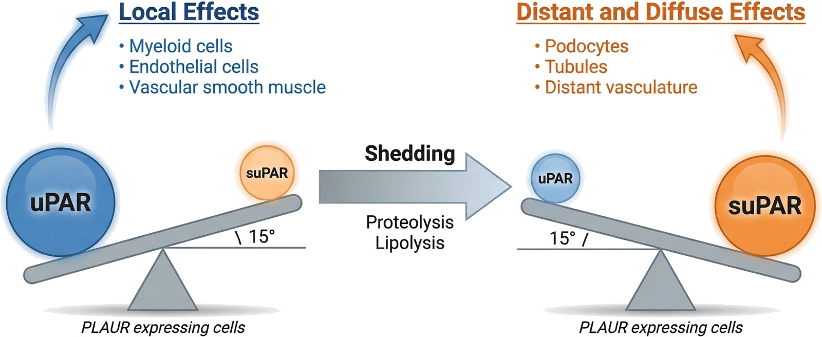
Cutting the receptor loose does not simply move it somewhere else. It removes the one thing that made the receptor useful, which is a location. What is left behaves differently, in two ways that pull in opposite directions.
The first is subtraction. A free receptor in solution still binds urokinase, but it does so nowhere in particular. A recombinant soluble receptor acts as a scavenger, taking urokinase away from cells that would otherwise dock it and dampening the plasminogen cascade rather than accelerating it (Wilhelm, FEBS Lett 1994; Behrendt, FEBS Lett 1996). In paroxysmal nocturnal hemoglobinuria, where plasma suPAR runs high, the excess competes with the membrane receptor on normal neutrophils and suppresses fibrinolytic activity at their surface (Ninomiya, Int J Hematol 1997). The soluble form works against the very thing the anchored form exists to do.
The second is addition, and it is the one that injures. The binding surfaces keep working when the anchor goes; they simply lose their address. Circulating suPAR engages αvβ3 integrin together with RAGE on podocytes, cells that never expressed the receptor, and that complex is required for everything that follows: Src phosphorylation, Rac1 activation, TRPC6 trafficking to the cell surface, reactive oxygen production. Plasma from a patient with recurrent focal segmental glomerulosclerosis activates podocytes in exactly this way, and a suPAR-neutralizing antibody blocks it (Kim, Biochim Biophys Acta Mol Basis Dis 2021). Formyl peptide receptors and β-arrestin-1 are part of the same machinery (Kim, Cells 2024).
So the two are not one molecule in two places. Membrane uPAR acts on the cell that made it, where its job is to aim. Soluble suPAR acts on cells that had no part in making it, where there is nothing to aim at. That is the difference between a tool and an injury, and it is the frame the lab's own 2026 review sets out: one gene product, compartmentalized, whose local and distant effects are not the same biology (Hayek, Circ Res 2026).
How suPAR is measured
Two very different technologies produce a number called suPAR, and they do not produce the same number. Which one was used decides whether a level can be compared with anything.
The reference method is an enzyme-linked immunosorbent assay, or ELISA, which catches the protein between a pair of antibodies and reports a concentration. Two of them are in common use and even they are not interchangeable: measured on the same samples they correlate at 0.75, and one reads about 50% higher than the other (Vasbinder, J Nephrol 2023). Some of that is the sugar chains the protein carries, since stripping them raises the measured concentration by about half (Montecillo, J Appl Lab Med 2025). A threshold therefore belongs to the assay it was derived on.
The other route is large-scale proteomics, where suPAR is one of thousands of proteins measured at once on an aptamer or antibody-pair platform. Those report relative abundance, normalized within a batch, rather than a concentration, and they track the ELISAs only loosely: a correlation of 0.29 between an aptamer platform and an ELISA in one cohort, and under 0.6 between a proximity-extension platform and an ELISA in another. The consequences are not academic. In the same participants, the ELISA measure separated cardiovascular death with a C-statistic of 0.74 against the aptamer platform's 0.57, and the aptamer measure lost the association with diabetes altogether (Vasbinder, J Nephrol 2023). Proteomics is what makes a study of tens of thousands of people possible, and it ranks risk within a cohort. But it cannot hand a patient a number, and a value from it cannot be read against a published cut-off. When the question is what someone's level is, the answer comes from an ELISA.
What a level means in a healthy person
In someone who is well, suPAR is not zero. It is a set-point, and the set-point tracks how that person has lived.
Measurable since an assay was developed for it in 1995 (Mizukami, Blood 1995; Brünner, APMIS 1999), it runs between roughly 2 and 4 ng/mL. Never-smokers in one study ran a median of 1.9 (Eugen-Olsen, Eur J Clin Invest 2016); participants of the Multi-Ethnic Study of Atherosclerosis who were free of cardiovascular disease, 2.5 (Hindy, J Clin Invest 2022); the Danish general-population cohorts, older and including smokers, a mean of 3.5 in men and 3.9 in women (Haupt, Biomark Insights 2014) and a median of 4.0 overall (Eugen-Olsen, J Intern Med 2010). There is no single normal range: the value depends on who is being measured, and on the assay used to measure them.
In 5,538 participants of Inter99, a Danish general-population study, suPAR was higher with age, in women, with lower socioeconomic position, with smoking in a dose-dependent way, with a poorer diet, with less physical activity and with higher body mass index (Haupt, Biomark Insights 2014). It also moves: among participants measured again five years later, those who stopped smoking, improved their diet or became more active saw smaller rises than those who did not (Haupt, Immun Ageing 2019).
That is the distinction that makes it useful, and it has a technical name: suPAR is not an acute-phase reactant. C-reactive protein and the other proteins a clinician reaches for when inflammation is suspected are made by the liver on demand, climb within hours of an infection, an operation or a heart attack, and fall when that resolves. suPAR barely registers those events (Rasmussen, Front Immunol 2021). What it reports is slower, closer to the accumulated load of immune activation a person is carrying, which is why one measurement in a well person forecasts, years ahead, what will happen to their kidneys and their arteries (Thunø, Dis Markers 2009).
Two things do move it quickly. Smoking is one: stopping brings the level down within four weeks, while C-reactive protein does not budge (Eugen-Olsen, Eur J Clin Invest 2016). RNA viruses are the other. SARS-CoV-2 raises suPAR in infected animals and in patients, and the rise is enough to injure the filtering cells of the kidney (Wei, Nat Commun 2023); levels run high in HIV as well, and stay high under treatment that suppresses the virus (Hoenigl, Clin Infect Dis 2019).
Three diseases that arrive together
A high suPAR predicts poor outcomes across a striking range of human illness. Working alongside other groups, we have spent a decade asking why.
Clinical, genetic and experimental evidence now place suPAR inside the biology of cardiovascular, kidney and metabolic disease rather than alongside it. The genetic case rests on a common variant in PLAUR that raises circulating suPAR and, in Mendelian randomization, tracks causally with atherosclerosis (Hindy, J Clin Invest 2022). Those three rarely travel alone: nearly 90% of American adults are at stage 1 or higher of cardiovascular-kidney-metabolic syndrome, and the risk of death climbs with every stage (Cheema, Am Heart J Plus 2026). Conventional risk factors do not fully account for that clustering, since patients whose glucose, lipids and blood pressure are all controlled still progress, which implies something running between the compartments (Hayek, Circ Res 2026). suPAR may be part of what connects them.
Where the lab has followed it:
- suPAR and kidney diseasePredicting chronic kidney disease before filtration falls, the APOL1 mechanism, and injury to the filtering cells themselves.
- suPAR and cardiovascular diseaseHuman genetics, Mendelian randomization and mouse models placing suPAR upstream of atherosclerosis rather than alongside it.