A high suPAR precedes symptoms by years
The first measurements were taken in people who felt well and whose arteries had not yet declared themselves. That is where a marker of risk earns its keep.
In 5,406 participants of the Multi-Ethnic Study of Atherosclerosis, all free of known cardiovascular disease when they enrolled, we measured suPAR and followed them for a median of 15 years. Coronary artery calcium, which a scan scores and which tracks how much atherosclerosis has accumulated, grew faster where suPAR was higher: it rose 103% over about two and a half years in participants below 2.0 ng/mL, against 229% in those above 3.0. Events followed the same order, 2.8 per 1,000 person-years in the lowest category and 9.0 in the highest, and each doubling of suPAR carried 1.46 times the risk after adjustment for the established risk factors and for kidney function (Hindy, J Clin Invest 2022).
The pattern had first appeared outside any clinic. In 2,602 Danish adults of the MONICA10 study, one baseline measurement predicted cardiovascular disease, diabetes, cancer and death over the following decade, independently of C-reactive protein (Eugen-Olsen, J Intern Med 2010), and adding it to the Framingham Risk Score reassigned people out of the intermediate band that score leaves unresolved (Lyngbæk, Int J Cardiol 2013). A protein that forecasts a heart attack ten years ahead in someone who feels well is either reporting on a process already under way, or taking part in it.
Three ways to ask whether suPAR causes atherosclerosis
No cohort, however large, can tell those two apart. Genetics and an animal can.
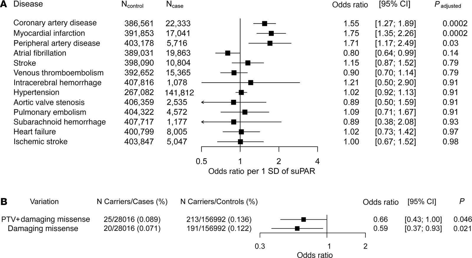
We measured suPAR in 12,937 people across four cohorts, searched the genome for the variants that set a person's level, and replicated the result in 12,177 more. Two common missense variants in PLAUR, the gene encoding the receptor, stood out, and only one of them, rs4760, actually raised secreted suPAR when it was expressed in human cells and in mice. A variant is inherited at random and independently of how a person lives, which makes it a natural experiment: in the UK Biobank, genetically predicted suPAR was associated with coronary artery disease, myocardial infarction and peripheral artery disease, and with none of the ten other cardiovascular conditions tested (Hindy, J Clin Invest 2022).
The mirror image holds. Across more than 280,000 exomes, people carrying rare damaging variants in PLAUR, the kind that should leave them with less of the protein, had 41% lower odds of ischemic heart disease (Hindy, J Clin Invest 2022). Two genetic approaches, pointing opposite ways for opposite reasons, agree.
Then the animal. Mice engineered to overexpress suPAR and normal mice of the same strain were given the same treatment to induce atherosclerosis and the same diet, and their cholesterol levels were indistinguishable. Their plaques were not: plaque volume at the aortic root averaged 1.55 mm³ against 0.90, the dead core inside it 0.18 mm³ against 0.05, and macrophages covered 47% of the plaque against 28% (Hindy, J Clin Invest 2022). The transgenic mice carry suPAR at concentrations no person reaches, so what the experiment fixes is the direction of the effect and not its size in humans. Raising suPAR alone, with cholesterol held equal, is enough to make the plaque worse.
Not another C-reactive protein
C-reactive protein is the inflammation test already in use, so the fair question is what a second one adds. They are not reporting the same process, and they do not behave alike when the same questions are put to them.
Put through the genetic test above, C-reactive protein does not survive it. Variants that raise a person's level for life, by as much as 30% per copy, carry no excess coronary disease: across 47 studies and 194,418 participants, the risk ratio for genetically raised C-reactive protein was 1.00 (Wensley, BMJ 2011), and a general-population study of more than 40,000 people reached the same conclusion (Zacho, N Engl J Med 2008). Measured levels do predict events, and the genetics say the protein is not what produces them. The same kind of instrument applied to suPAR returned 1.55 for coronary artery disease. One of them reports the fire. The other helps set it.
The difference starts with where each comes from. C-reactive protein is an acute-phase protein, made by the liver on demand when interleukin-6 rises, so it tracks the infection or injury of the moment and falls when that resolves. suPAR is not an acute-phase reactant at all. It is shed from the surface of immune cells themselves and moves slowly: no variation across the day, about 10% variation within a person (Thurison, Clin Chim Acta 2015), and little answer to the insult of the hour (Rasmussen, Front Immunol 2021). The contrast shows inside a single admission. In 296 patients treated with angioplasty for a heart attack, C-reactive protein rose and fell over the following days while suPAR held steady, and over the next five and a half years suPAR predicted death and a second infarction while C-reactive protein predicted neither (Lyngbæk, Am J Cardiol 2012). The few things that do raise it quickly are specific: smoking, and infection with an RNA virus such as SARS-CoV-2 or HIV (Eugen-Olsen, Eur J Clin Invest 2016; Wei, Nat Commun 2023). One measurement of a stable protein carries further than one measurement of an unstable one.
They also do not pick out the same people. In 1,126 randomly sampled middle-aged adults, suPAR was associated with the amount of calcium in the coronary arteries and high-sensitivity C-reactive protein was not (Sørensen, Atherosclerosis 2014). In 1,703 patients with established coronary disease, suPAR predicted cardiovascular death and infarction independently of C-reactive protein, troponin, natriuretic peptide and kidney function (Nikorowitsch, J Am Heart Assoc 2020), and in 500 African Americans with type 2 diabetes it was the only one of three markers still associated with death when it, coronary artery calcium and high-sensitivity C-reactive protein were entered together (Hayek, J Am Heart Assoc 2018). Neither test substitutes for the other, and a risk score can carry both (Desai, Am J Cardiol 2023).
The trial that put the two side by side is ours. In BARI 2D, 2,277 patients with type 2 diabetes and coronary artery disease had both markers measured at entry and 1,978 again a year later, under intensive guideline-directed care. High-sensitivity C-reactive protein fell by roughly a third over that year, from a median of 2.07 to 1.30 mg/L. suPAR did not move in any treatment arm. It went on predicting death, myocardial infarction and stroke at 1.40 times the risk per standard deviation, undiminished by adjustment for C-reactive protein, and it still separated risk among the patients whose C-reactive protein had normalized: classified by both markers together, five-year event rates ran from 7.1% to 21.6% (Ismail, Diabetes Care 2026). A patient whose inflammation looks treated by the test in use can still be carrying it.
Persistently high suPAR primes the monocyte
Atherosclerosis is built by monocytes that leave the blood and settle in the wall of the artery. Hold suPAR high for long enough and those cells are already altered before they get there: quicker to move, and set to overreact when something provokes them.
In mice with high suPAR levels but with no atherosclerosis induced, the aorta was already changed. It secreted more CCL2, the chemokine that calls monocytes in, and held twice as many monocytes. Those monocytes displayed more of the receptors that answer that call, CCR2 and CX3CR1, and in a migration assay more of them crossed the membrane, with or without added CCL2 (Hindy, J Clin Invest 2022).
What sustained exposure changes is how the cell answers the next insult. Macrophages grown from bone marrow and pre-treated with suPAR are not inflamed by suPAR itself. Provoked afterwards, they produce far more tumor necrosis factor-α, interleukin-1β and interleukin-6, and mount a much larger NLRP3 response, than macrophages never exposed to it. In animals, the same sustained elevation expanded inflammatory CCR2-positive monocytes and macrophages in the heart, the spleen and the blood at once, and worsened diastolic function (Singh, bioRxiv 2026). suPAR is not the alarm. It is what leaves the alarm set too loud.
This is the anchored receptor's own job, turned on the vessel. Cut loose, the protein that aims a monocyte's enzymes instead acts on cells that never belonged to it, and what it leaves behind is a monocyte readier to move and readier to inflame (Hayek, Circ Res 2026). Whatever a patient's lipids are doing, this runs on a separate track.
What a level says in someone already ill
The people whose risk most needs sorting are rarely well. In them, suPAR adds to the tests already in use rather than repeating them.
Start with the numbers themselves. In people free of cardiovascular disease the median is about 2.5 ng/mL, and risk begins to separate above 3.0 (Hindy, J Clin Invest 2022). Patients with heart failure run a median of 3.4 (Hayek, J Card Fail 2023), and those hospitalized for worsening heart failure with diabetes 4.7 (Ismail, J Card Fail 2026). Those thresholds are not portable. They differ by sex: women run about 10% higher than men, enough that a single cut-off misclassifies, and with sex-specific ones, 4.4 ng/mL in women against 3.2 in men, the same level carries the same risk in either (Mehta, J Am Heart Assoc 2020). They differ by assay too. Every figure on this page comes from the same ELISA, and a value produced by a different method cannot be read against them.
In heart failure it adds to the test already in use. Among 1,116 such patients followed for a median of 6.2 years, each doubling of suPAR carried 2.30 times the risk of death after adjustment for B-type natriuretic peptide, and it sharpened discrimination whether the ejection fraction was reduced or preserved (Hayek, J Card Fail 2023). It separates risk most sharply where least can be done about it: among 406 patients with preserved ejection fraction in the TOPCAT trial, five-year cardiovascular death, cardiac arrest or heart failure hospitalization ran 44% in the highest third against 14% in the lowest, independently of natriuretic peptide (Hutten, ESC Heart Fail 2025).
The same holds outside the coronary artery, and outside the heart. In 5,810 patients undergoing cardiac catheterization suPAR identified peripheral arterial disease and the events that followed it (Samman, Atherosclerosis 2017), the territory the genetics flagged. And the excess this page opens with is measurable in the same way: people with chronic kidney disease carry cardiovascular risk beyond what their measured risk factors explain (Go, N Engl J Med 2004), and in 4,994 of them about two thirds of suPAR's association with cardiovascular death ran directly rather than through kidney function (Sommerer, Kidney Int Rep 2023). The two diseases look less like one causing the other than like both answering to something shared.
Inflammation the treatment does not reach
Three randomized trials have now measured suPAR before and after a therapy that works. None of them moved it.
BARI 2D, above, is the first: a year of intensive glycemic and revascularization strategy left suPAR where it started, in every arm. The other two tested drugs that improve outcomes. Spironolactone left suPAR unchanged at one year in the TOPCAT patients (Hutten, ESC Heart Fail 2025), and sotagliflozin, which cut heart failure events by a third in SOLOIST-WHF, lowered it no more than placebo did, with the same benefit at every suPAR level (Ismail, J Card Fail 2026).
These are useful negative results: those drugs help through something else, and this particular risk is still sitting in patients already treated to target. Inflammation itself is a proven cardiovascular target, since blocking interleukin-1β lowered events without changing lipids at all (Ridker, N Engl J Med 2017). A pathway that nothing in current use reaches is therefore a candidate for a drug, and not only for a test.
It is not that the level cannot be moved. In a randomized study of 48 smokers, four weeks of stopping brought suPAR down to the range of people who had never smoked, while C-reactive protein did not respond at all (Eugen-Olsen, Eur J Clin Invest 2016), and the same is seen over years in people who improve their diet and activity (Haupt, Immun Ageing 2019). What does not move it is the pharmacology, which is precisely why the risk it marks is still there after the prescription.
What it would take to treat it
Nothing in clinical use lowers suPAR. But the first antibodies built to block it are in patients now, which turns the question from a hypothetical into a trial.
The antibody that blocks suPAR prevents kidney injury in mice (Hayek, N Engl J Med 2020), and antibodies of that kind have left the animal work behind: one has completed a phase 1 safety study in healthy volunteers and is now in a randomized, placebo-controlled trial in patients with glomerular kidney disease, with anti-suPAR therapy under active clinical evaluation (Yang, Front Endocrinol (Lausanne) 2026). None has yet been tested against atherosclerosis or any cardiovascular endpoint, so for the moment this evidence supports selection rather than treatment: identifying the patients whose remaining risk is of this kind, in a population whose glucose, blood pressure and lipids are already handled and whose events keep happening anyway.
The reason to keep at it is that the artery is not the only place this shows. The same protein predicts kidney decline in the same people, injures the filtering cells of the kidney directly, and marks cardiovascular risk in survivors of breast cancer, among whom the highest quartile carried three times the risk of the lowest over 13 years (Yadalam, J Am Heart Assoc 2025). Nearly 90% of American adults sit at some stage of cardiovascular-kidney-metabolic syndrome (Cheema, Am Heart J Plus 2026), and a factor that acts in all three of its compartments is a candidate for what connects them.
Read next: